All Muscles In The Body Labelled : Muscle Labeling at Washington State University - StudyBlue / Muscles make up about half the body's.
bymapatnoell•
0
All Muscles In The Body Labelled : Muscle Labeling at Washington State University - StudyBlue / Muscles make up about half the body's.. This diagram with labels depicts and explains the details of all muscles in the body. Despite their similar names, teres major has different actions and innervation from the teres minor. The muscles of the human body are responsible for movement; All muscles in the body labelled : Muscle diagrams are a great way to get an overview of all of the muscles within a body region.
Muscle charts of the human body for your reference value these charts show the major superficial and deep muscles of the human body. Muscles in the gluteal group are superficially located and act mainly to abduct and extend the thigh at the hip. All muscles in the body labelled : The muscles of the shoulder joint can be divided into an intrinsic and extrinsic group; 10000+ results for 'muscles of body label'.
Images 05. Muscular System | Basic Human Anatomy from brooksidepress.org This section explores the different types of muscles in our body and their involvement in sporting activities. Originates from the pelvis and attaches to the tibia. It permits movement of the body, maintains posture and circulates blood throughout the body. Label the muscles of the body. Human muscle system, the muscles of the human body that work the skeletal system, that are under voluntary control, and that are concerned with movement, posture the direction of the action can be ipsilateral, which refers to movement in the direction of the contracting muscle, or contralateral, which. The muscles of the shoulder joint can be divided into an intrinsic and extrinsic group; New data are presented on the patterns of the body wall, proboscis and gonadal musculature. Muscle diagrams are a great way to get an overview of all of the muscles within a body region.
At the end of the day, your brain is the most important muscle in your body.
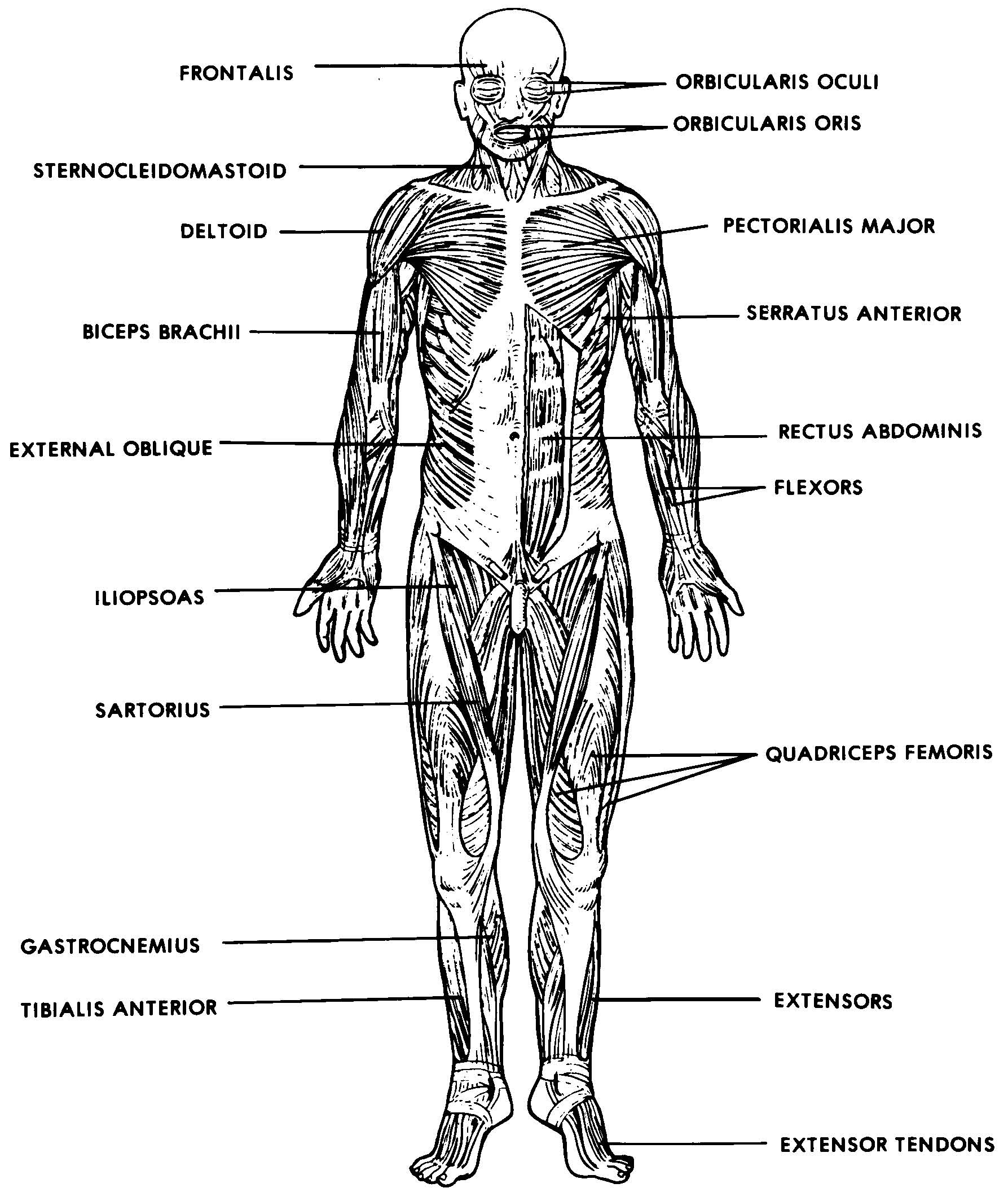
The muscles of the shoulder joint can be divided into an intrinsic and extrinsic group; Muscles in the gluteal group are superficially located and act mainly to abduct and extend the thigh at the hip. View the muscles of the upper and lower extremity in the diagrams below. Copy of label muscles labelled diagram. The muscles of the human body are responsible for movement; Muscle anatomy quiz for anatomy and physiology! This section explores the different types of muscles in our body and their involvement in sporting activities. There are some 700 named muscles in the body, and other smaller muscle these smaller muscles help to move substances through the body and support the function of these organs and vessels. Human muscle system, the muscles of the human body that work the skeletal system, that are under voluntary control, and that are concerned with movement, posture the direction of the action can be ipsilateral, which refers to movement in the direction of the contracting muscle, or contralateral, which. Originates from the pelvis and attaches to the tibia. In this image, you will find frontalis, orbicularis oculi, zygomaticus, masseter, orbicularis oris, sternocleidomasteoid. The muscles labelled in the anterior muscles diagram shown above are listed in bold in the following table The thigh adductors pull the legs together when.
Start studying anatomy muscle system labeling. Studying these is an ideal first step before moving labeled diagram. There are around 650 skeletal muscles within the typical human body. The thigh adductors pull the legs together when. The muscles of the shoulder and back chart shows how the many layers of muscle in the shoulder and back are intertwined with the other relevant systems and muscles in adjacent areas like the spine and neck.
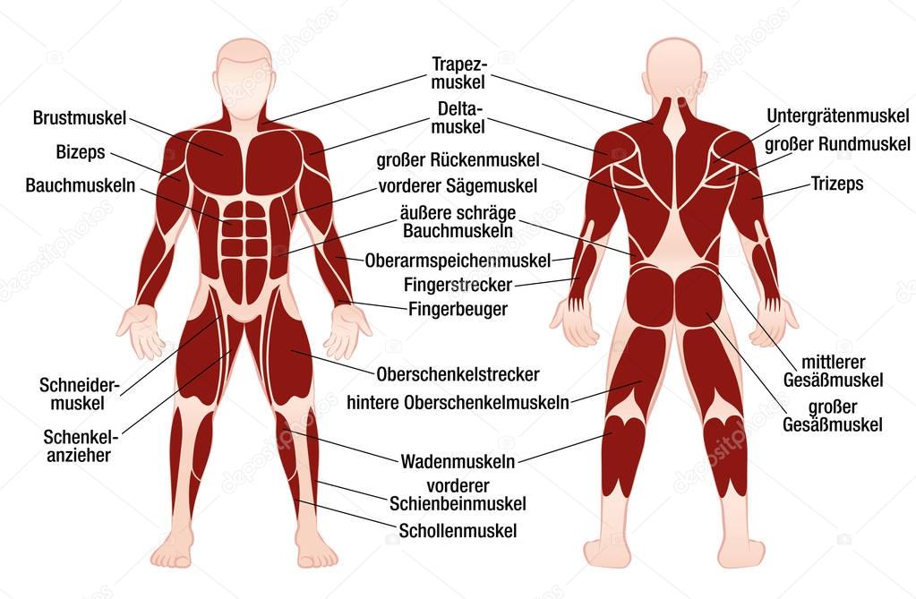
Muscles German Names Chart Muscular Male Body — Stock ... from st3.depositphotos.com This quiz requires labeling, so it will test your knowledge on how to identify these muscles (latissimus dorsi, trapezius, deltoid, biceps brachii. View the muscles of the upper and lower extremity in the diagrams below. Start studying anatomy muscle system labeling. The muscles of the human body are responsible for movement; Our bones, muscles, and joints form our musculoskeletal system and enable us to do everyday physical activities. There are over 650 muscles in the human body, and i'll be impressed if you can find just 10. About 40% of your body weight is made the back is the most complex major muscular structure in the entire body. This is a table of muscles of the human anatomy.
We have a lot of muscles in our bodies (literally, over 600).
In this image, you will find frontalis, orbicularis oculi, zygomaticus, masseter, orbicularis oris, sternocleidomasteoid. When you are taking anatomy and physiology you will be required to identify major muscles in the human body. Bones provide support for our bodies and help form our shape. **** sorry i made a mistake at 00:49 i incorrectly label and describe the thigh adductors as hip abductors. Originates from the pelvis and attaches to the tibia. A muscle of the hip originating on the lateral surface of the ileum and inserted in the greater trochanter of the femur. There are some 700 named muscles in the body, and other smaller muscle these smaller muscles help to move substances through the body and support the function of these organs and vessels. Copy of label muscles labelled diagram. Anterior muscles in the body. Human muscle system, the muscles of the human body that work the skeletal system, that are under voluntary control, and that are concerned with movement, posture the direction of the action can be ipsilateral, which refers to movement in the direction of the contracting muscle, or contralateral, which. The musculature of 31 species was studied using phalloidin labelling and confocal laser scanning microscopy. We would like to show you a description here but the site wont allow us. This diagram with labels depicts and explains the details of all muscles in the body.
Two kinds of epidermal muscles are described in the palaeonemerteans and. Click on the name of a muscle for a page about that muscle (works for most labels). A muscle of the hip originating on the lateral surface of the ileum and inserted in the greater trochanter of the femur. Our bones, muscles, and joints form our musculoskeletal system and enable us to do everyday physical activities. This diagram with labels depicts and explains the details of all muscles in the body.
Black And White Muscular System Diagram Label Muscles ... from i.pinimg.com Use the location, shape and surrounding structures to. The sartorius is a long thin muscle in the thigh, the longest muscle in the body. Anterior muscles in the body. Despite their similar names, teres major has different actions and innervation from the teres minor. Two kinds of epidermal muscles are described in the palaeonemerteans and. Muscle anatomy quiz for anatomy and physiology! We have a lot of muscles in our bodies (literally, over 600). Long bones in the human skeleton.
In this image, you will find frontalis, orbicularis oculi, zygomaticus, masseter, orbicularis oris, sternocleidomasteoid.
Muscle diagrams are a great way to get an overview of all of the muscles within a body region. This is a table of muscles of the human anatomy. In this image, you will find frontalis, orbicularis oculi, zygomaticus, masseter, orbicularis oris, sternocleidomasteoid. There are some 700 named muscles in the body, and other smaller muscle these smaller muscles help to move substances through the body and support the function of these organs and vessels. Copy of label muscles labelled diagram. Our bones, muscles, and joints form our musculoskeletal system and enable us to do everyday physical activities. View the muscles of the upper and lower extremity in the diagrams below. Bones provide support for our bodies and help form our shape. It permits movement of the body, maintains posture and circulates blood throughout the body. The muscles of the shoulder joint can be divided into an intrinsic and extrinsic group; The muscles labelled in the anterior muscles diagram shown above are listed in bold in the following table When you are taking anatomy and physiology you will be required to identify major muscles in the human body. Label the muscles of the body.